
Core advantages
Observe targets directly on the membrane with less loss from elution and transfer
Reduce morphology changes caused by repeated handling
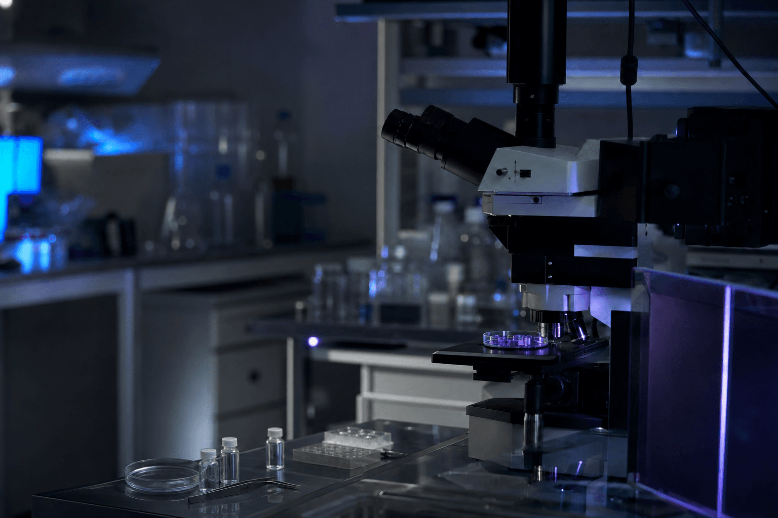
Connect bright-field microscopy, morphology and compatible fluorescence methods
Applicable samples and scenarios
Evaluation examples: cerebrospinal fluid, pleural or ascitic fluid, urine, water, culture media and other method-compatible liquid samples
Evaluation examples: cells, bacteria, fungi, parasite eggs and other small particles
Bright-field microscopy, morphology and compatible fluorescence workflows
Workflow
Step 1Sample pretreatment
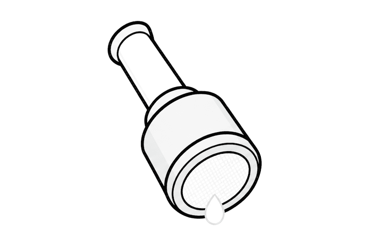
Prepare the sample
Select pretreatment from the sample matrix and target characteristics so particles are evenly dispersed.
Step 2Enrichment
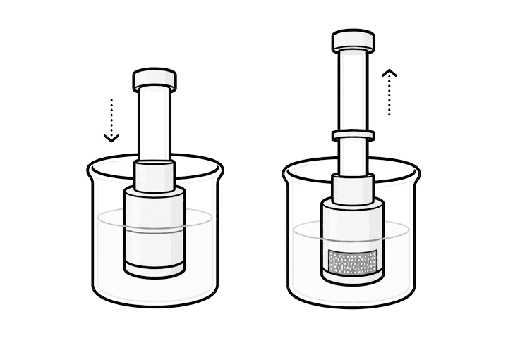
Filter and concentrate
Pass the sample through the shadowless membrane so target cells, microbes or particles are retained in a small surface area.
Step 3Slide preparation
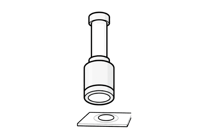
Prepare directly on the membrane
Move the enriched membrane directly into slide preparation, clearing or staining without first eluting targets back into liquid.
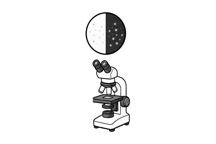
Step 4Microscopy
Microscopy and fluorescence
Observe concentrated targets by bright-field or fluorescence microscopy with more targets in each viewing area.
Recommended product configuration
Shadowless membrane concentration sampler and slide-preparation device
The core consumable for filtration enrichment, membrane preparation and direct microscopic observation.
Data and research evidence
On-membrane
Observe after concentration
Targets remain concentrated in a small membrane area, increasing target density within each observation field.
No elution
Reduce recovery loss
Compatible workflows do not require targets to be eluted from the membrane back into liquid first.
Fewer transfers
Simpler sample movement
Fewer tube changes, recovery steps and repeat slide preparations reduce losses of low-abundance targets.
Less disturbance
Preserve morphology
Less repeated centrifugation, resuspension and transfer helps preserve cell and particle morphology.
Get a solution
Bring more of the target into your detection workflow
Tell us your sample, target and current detection method. We will recommend the membrane route, consumables and workflow connection.
Get a microscopy and fluorescence solution