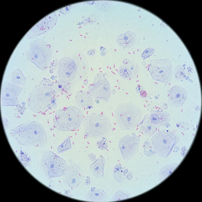
Centrifugation / embedded adsorption
The sandwich cup is used in the operation, with centrifugal action and embedded adsorption enriching target material at the ultra-thin membrane surface.
 Get a solution
MEMBRANE ENRICHMENT PLATFORM
Centrifugation / embedded adsorption or filtration / retention concentrates targets and retains them at the membrane surface. Typical workflows reach about 20–100× enrichment depending on sample volume and process conditions, and matched clearing reduces the light scattering caused by air in the membrane pores, before morphology, fluorescence, nucleic-acid or other detection.
INTERACTIVE PRINCIPLE
After the required handling, the sample enters centrifugation / embedded adsorption or filtration / retention. Targets concentrate at the membrane surface before morphology, fluorescence, nucleic-acid or other analysis.
The sample enters one of two membrane-enrichment routes
After the required sample handling, bring the sample into the centrifugal sandwich cup or filtration device.
Centrifugation / embedded adsorption drives targets toward the ultra-thin membrane; filtration lets carrier pass while targets are retained. The routes are selected independently, but both concentrate targets at the membrane surface.
The enriched membrane connects directly to downstream detection. Compatible microscopy workflows can observe targets directly, while PCR / NGS workflows can enter lysis, amplification or library preparation without first eluting targets back into liquid; each method keeps its required downstream steps.
The sandwich cup is used in the operation, with centrifugal action and embedded adsorption enriching target material at the ultra-thin membrane surface.
The sample passes through the membrane, where target material is retained and enriched; membrane transfer may be used.
The mechanisms differ, but both routes concentrate targets and retain them at the membrane surface.
Principle schematic · time is accelerated; samples and operating conditions follow the method used.
Drag to rotate · Ctrl / ⌘ + scroll to zoom
Tap “Explore model”, then drag to rotate and pinch to zoom
Loading the 3D demo…
Drag to rotate · Ctrl / ⌘ + scroll to zoom
Tap “Explore model”, then drag to rotate and pinch to zoom
Loading the 3D demo…
The sample enters one of two membrane-enrichment routes
After the required sample handling, bring the sample into the centrifugal sandwich cup or filtration device.
Centrifugation / embedded adsorption drives targets toward the ultra-thin membrane; filtration lets carrier pass while targets are retained. The routes are selected independently, but both concentrate targets at the membrane surface.
The enriched membrane connects directly to downstream detection. Compatible microscopy workflows can observe targets directly, while PCR / NGS workflows can enter lysis, amplification or library preparation without first eluting targets back into liquid; each method keeps its required downstream steps.
The sandwich cup is used in the operation, with centrifugal action and embedded adsorption enriching target material at the ultra-thin membrane surface.
The sample passes through the membrane, where target material is retained and enriched; membrane transfer may be used.
TECHNOLOGY ROUTES
The routes are chosen independently by sample, target and downstream method; each route page explains its principle, workflow and products.
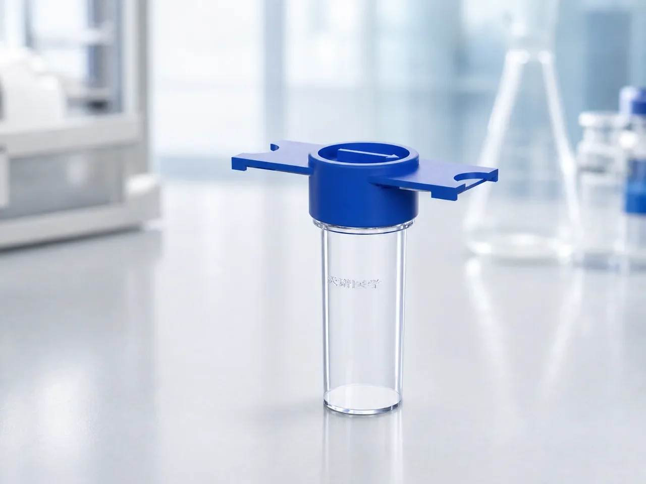
ROUTE A · Centrifugal action
Centrifugation and embedded adsorption concentrate dispersed targets at an ultra-thin membrane surface before clearing, slide preparation, staining and digital review.
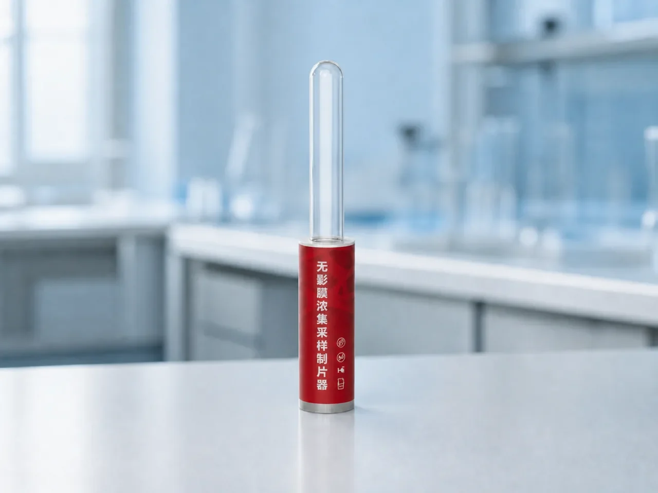
ROUTE B · Direct filtration
Filtration and retention concentrate small particles from a larger liquid sample onto one membrane, ready for microscopy, fluorescence or PCR / NGS preprocessing.
FAQ
Questions and answers last reviewed October 28, 2026.
ENRICHMENT FIRST · DETECTION NEXT
The enriched membrane interface can connect to morphology, fluorescence, nucleic-acid and other methods as needed. Our technical team can explain how the platform fits the existing workflow and which product system carries it forward.